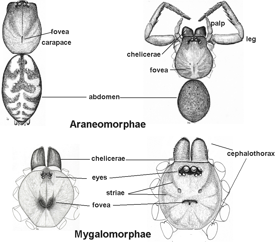
Spiders have two main parts to their body - the cephalothorax and the abdomen. The abdomen is the posterior portion of the spider; also called the opisthosoma.

A hardening of the cuticle of the dorsal, lateral or ventral abdomen that forms a shield or plate which is often shiny and pitted.
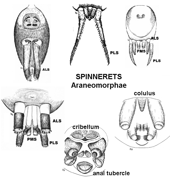
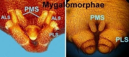
Anterior lateral spinnerets. ALS are absent in mygalomorphs with 4 spinnerets, but are present in all araneomorphs. In the Prodidomidae: Molylcriinae, the ALS are grossly enlarged and positioned greatly forward/anterior to the other spinnerets.

Anterior median spinnerets. Present as actual spinnerets only in the Liphistiidae (mygalomorph-like spiders with abdominal segmentation still evident). The cribellum in araneomorph spiders is a homologue of the AMS, and it may be reduced to a non-functional fleshy colulus or lost altogether.

A pair of roughly triangular, soft projections above the anus of all spiders. It is situated on the last abdominal segment, just posterior to (or behind) the spinnerets. Fecal matter is discharged from here.

At the front.

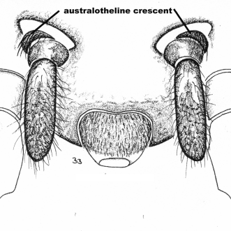
A crescent-shaped ridge of hair or sclerotised ridge locataed anterior to the PMS in some diplurid mygalomorphs. The ridge is isolated from surrounding cuticle by a narrow unsclerotised area.

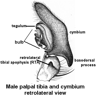
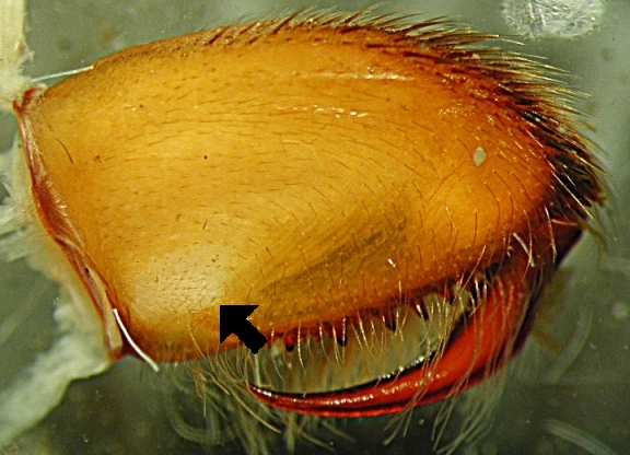
When the base of the cymbium is basally constricted into a low ridge or conical process.


Total body length is from the front of the chelicerae to the back of the abdomen, and does not include the spinnerets. Although body size may be considered a weak character, it can sometimes be very informative. For example, if your spider lies in the small to minute size group and is an adult, it is likely to be one of the 'micro' spiders which usually have the leg tarsi longer than the metatarsi, and this should be examined. Equally, few spiders belong to the extremely large group, and if you have such a spider then the character should be scored.
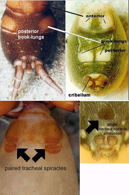
The large respiratory structures of spiders. In most araneomorph spiders, only the anterior book-lungs are present, and are visible as transverse slits in line with the female genital organ.

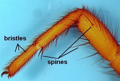
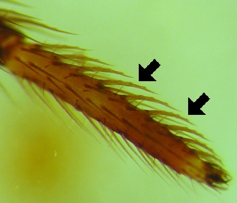
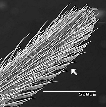
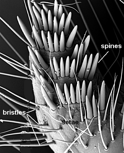
Bristles, setae and hairs are considered here to be different to spines, in that they are not movable by hydraulic pressure in the limb. Some long, slender and thick bristles may appear to be spines, but they cannot be raised and lowered. Bristles and setae tend to be tapered towards the tips and to be gently curved in one direction. Bristles are considered here to simply be thicker setae that tend to be longer and to taper much more gradually than the shorter setae. Hairs tend to be slender and abruptly tapering, and almost adherent to the cuticle of the leg, carapace or abdomen. See also spine(s).

A fine curved comb of hair located dorsally on metatarsus IV of cribellate spiders. The calamistrum can be one or more lines or a band, and is used to comb cribellate silk from the cribellum.
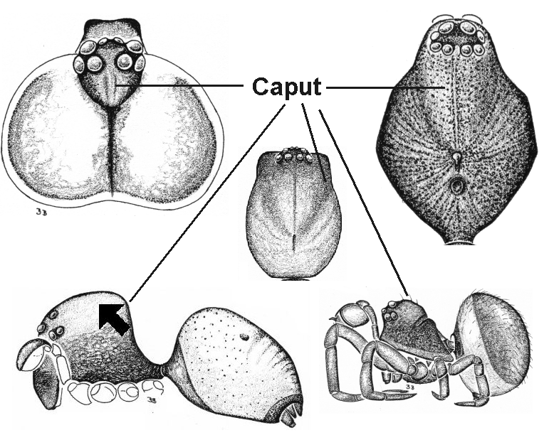
The portion of cephalothorax anterior to the fovea (when present). The caput is often higher set than the posterior portion of the cephalothorax, and is sometimes referred to as the thorax.
The carapace is the hard sclerotised dorsal surface of the cephalothorax.
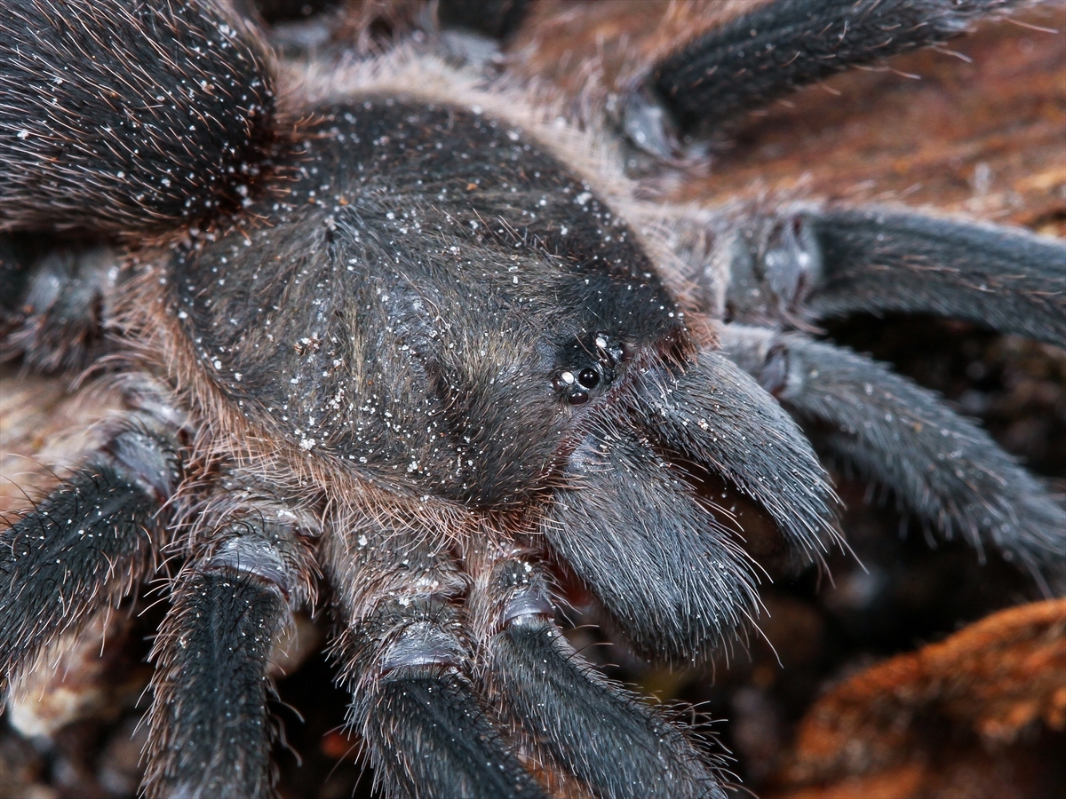
Spiders have two main parts to their body - the cephalothorax and the abdomen. The cephalothorax is the front half of the body from which the legs and chelicera (bearing the fangs) arise; also called the prosoma.
Two blunt lobe-like appendages on the front of the carapace from which the fangs arise ventrally.


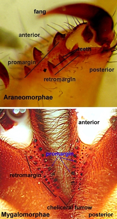
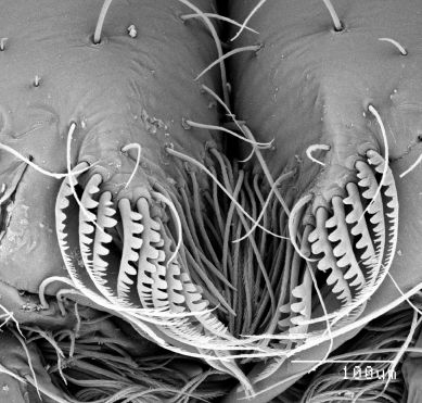
Pointed extensions of cuticle from the cheliceral margin against which the fang acts when it closes or retracts. Cheliceral teeth have no socket at the base and hence differ from peg teeth, spines and setae.

A sclerotised transverse plate, divided or entire, located between the carapace and chelicerae of some araneomorph spiders, e.g. Zodariidae. The chilum is not readily visible as the chelicerae are usually withdrawn, and hence this character should not be scored.
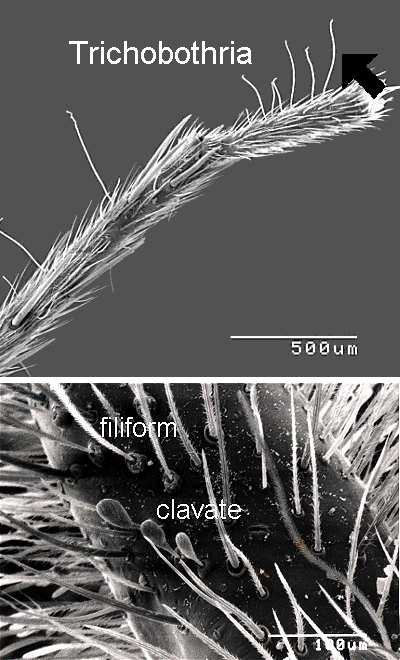
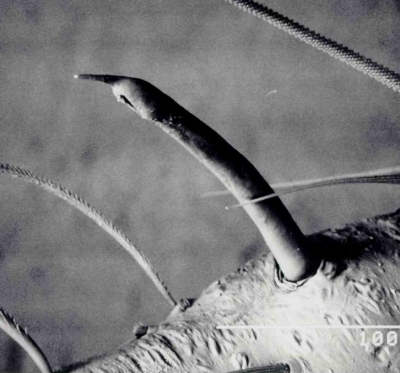
Paddle-like in shape, referring here to trichobothria in mygalomorphs (see trichobothria).

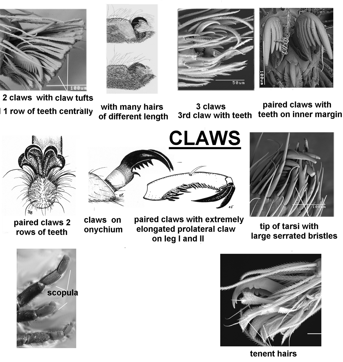
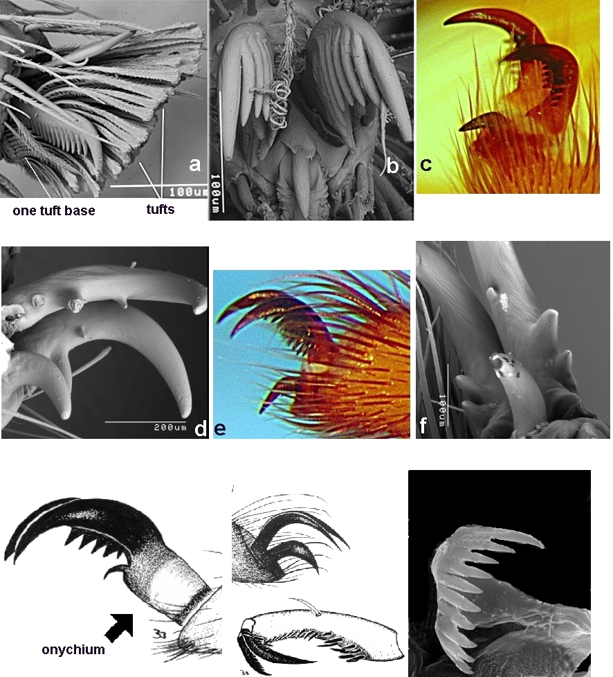
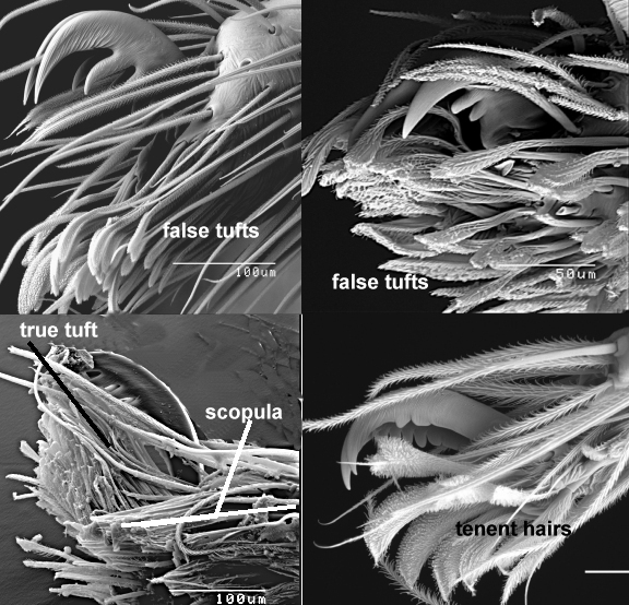
Here claw tufts are regarded as dense groups of hairs that arise from discrete pads between the outer surfaces of the claws and the cuticle of the lateral tarsus. The pads may be fused to the claws and tarsus. True tufts are under hydraulic control, and this can easily be seen under a dissecting microscope by compressing the tarsus with a pair of forceps. See also scopula or tenent hairs.


The most pleisiomorphic (or least modified) spiders, such as Liphistiidae, have three claws on each leg (a pair and a single) and one claw on the palp. The paired claws are larger and set high on the ovoid unsclerotised portion of the apical tarsus. Below this pair is a smaller unpaired claw. In hunting spiders and most tarantulas, e.g. Salticidae jumping spiders, the third claw is lost and instead, a dense pad of hairs called the claw tuft is present on each side of the paired claws. Most claws have ventral teeth, which appear as elongate triangular processes on the underside of the claw.
The least modified (pleisiomorphic) condition of the claws is the presence of a single row of teeth medially on the paired claws, third claw and palp claw. Modifications on that basic condition in the paired claws include an S-shaped row of teeth, a row of teeth on one or both outer faces of the claw, or no teeth on the claws at all. The latter condition occurs in the large claw-tufted tarantulas and brush-footed trapdoor spiders.
Two rows of teeth (one on each face) occur more diversely from the tiny armoured araneomorph Oonopidae and Orsolobidae, to the large mygalomorph Dipuridae and Nemesiidae. Establishing how many rows of teeth on the paired claws can be quite difficult, especially on small spiders and on claw-tufted spiders. It is best to place the spider with the ventral surface upwards and to examine the claws by holding the tarsi slightly past the verticle so that the ventral surface of the claws is parallel to the viewing plane. By slowly rocking the tarsus, the double row should be easily seen. In a quirk of evolution, the number of teeth on the claws of spiders varies between adult male and female of the same species. The female of some brush-footed trapdoor spiders (Barychelidae) may have few teeth in a single medial row, whereas the male of the same species may have two distinct rows of teeth. In some south-east Asian nemesiids, the female has two rows of teeth but the male has only one row.
If only a single row of teeth is present on the claws, the location of that row can have taxonomic importance. Sometimes the row is on the lateral faces of the claw, suggesting that it is either the precursor of the two rowed condition or it is a later stage in which one row was lost.
The palpal claw is typically present and has few teeth in a single row. In some groups, the claw may be obscured by hair, but evident when viewed by looking at the claw in line with the long axis of the tarsus. The claw is absent in some groups, but these are usually quite small spiders and the absence of such a tiny structure is difficult to establish. Rarely, the palpal claw is highly modified as is seen in the Hadrotarsinae (Theridiidae). The palpal claw is present in females and juvenile males; while in adult males it is transformed into a highly modified bulb used for the transfer of sperm to the female (see male palp).

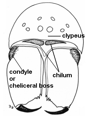
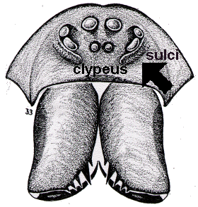
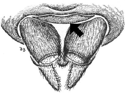
When the spider is viewed from the front, the clypeus is the space between the most forward set eyes (AME) and the edge of the carapace.
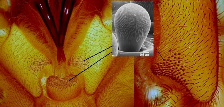
Situated immediately in front of the spinnerets on ventral posterior abdomen. The colulus is a hairy thumb-like lobe or wide plate (lacking spigots) that represents a non-functional cribellum. See also AMS.

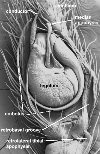
A process on the male palp which serves to support the tip and sometimes also the greater length of the emolus on its path.

Also called the 'cheliceral boss', the condyle is a small but distinctly raised mound on the outside of the chelicerae near its junction with the carapace. The condyle is best seen when spider viewed from the front.
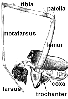
The first segment of the leg, only visible when spider is viewed from below. The coxae are typically rectangular in shape. The coxae of the palps are called maxillae (also gnathocoxae or endites). See leg segments.

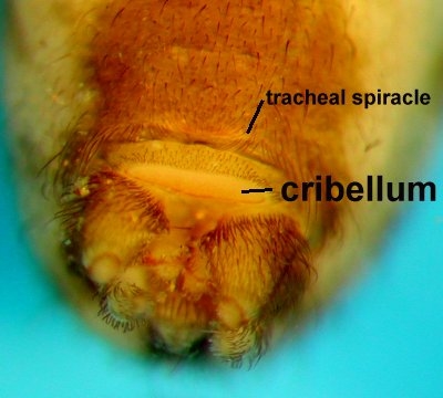
A transverse flat plate in front of the spinnerets on ventral posterior abdomen, that represents the transformed anterior spinnerets (AMS) in non-mygalomorph spiders. It is usually oval in shape, and in some groups may be difficult to see as it is often folded when preserved with only the edge visible. The cribellum is present in males but is non-functional. The bright blue cribellate silk used by net-casting spiders to adorn their web is extruded from the cribellum. The silk of other cribellate spiders may not be so blue, but they can produce what is known as hackled bands of silk.


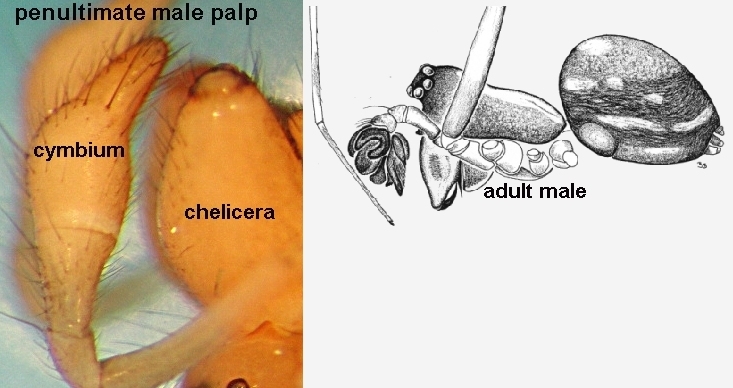
The terminal segment (or tarsus) of the male palp. In penultimate developmental stage of araneomorphs, the tarsus is swollen basally, and only becomes the cymbium in the final moult to adult. The term cymbium is usually used only for araneomorph palps; whereas it is simply referred to as the tarsus on mygalomorphs. See also basodorsal process.


When the action of the fang is across the body (see fang action).


The distributions provided here are taken from museum records and any other empirical information for that taxon. Some, however, are partially predictive. For example, a group which is known from the rainforest in north Queensland is likely to also be present in the rainforest of the Northern Territory. In some cases, better results will be obtained if the adjacent state is also scored. However, the Tasmanian fauna is well known at the subfamily level, and if the spider is from that state alone, that should be scored.
When viewed from the front angle, the lateral edges of the eye row are curved downwards. Eye row curvature is measured by a line drawn through the centers of the eyes in that row.
A modification of the claw on the male palp to become the palp bulb. Sperm is lifted from the sperm web into the sole opening of the bulb, which is located at the end of a spine-like tube, and then discharged into the female.


The form of female genitalia in araneomorph spiders in which the sperm is transferred to the female via an insemination duct, but fertilisation of the eggs occurs via a different duct, the fertilisation duct. An entelegyne epigyne may be slightly to very sclerotised externally and often has lateral teeth, media septa, and lateral lobes.
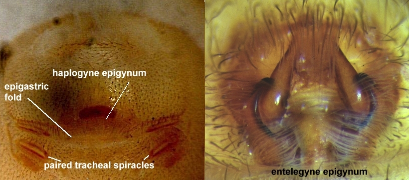
The external female genitalia. The term is usually used only with entelegyne araneomorph spiders. Sperm is transferred to the female epigyne by the male palp, whereby the male embolus is inserted into the female copulatory opening.

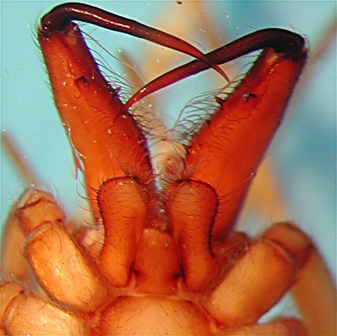
The sharp, curved tapering and highly sclerotised distal joint of the chelicerae. The venom gland passes through the fang and discharges from a small slit on the outer edge just before the pointed tip.
The direction in which the fangs are articulated and move when being used. The fang action can be transverse (across the body or diaxial), longitudinal (down the body, or paraxial) or diagonal and intermediate between the former two options. Fang action is related to, but not strictly correlated with, the direction in which the base of the chelicerae are articulated. In mygalomorphs, the action of the basal cheliceral segment is up and down to the front. This produces a striking action like that of a snake. Mygalomorphs must hold onto the surface that they are attacking when they strike. In all araneomorphs, the basal cheliceral segment is articulated to the side, so that the fangs pinck towards each other, and most araneomorphs can bite an object without having to touch or hold it.
Note that in some araneomorphs, the chelicerae are long and extended forwards (e.g. Tetragnathidae) and the fangs are long and set longitudinally (aligned with the body axis). However, as Patnick notes (pers. comm.), the action of the fang may be paraxial (longitudinal with the body axis) but the basal cheliceral segment is diaxial (across the body). Hence, the emphasis here is on the action of the fang.
The enlarged black (and often serrated) setae that form a line on the front face of the chelicerae, just beside the base of the fang. They are clearly visible with a binocular microscope when the spider is viewed from the front angle or from below. The setae are only present in females, and are most dramatic in the Corinnidae: Castianierinae. Also called cheliceral shield.
The third segment of the legs (after coxa and trochanter); or the first long segment of the legs and palp (counting from body out along leg). See leg segments.

Like a filament or wire; with similar width for the length of the structure; usually refering to a hair, seta or trichobothria. See also trichobothria.
A groove (longitudinal or transverse) or pit on the posterior half of the carapace. It is the external indication of the point where the sucking stomach is attached. In most mygalomorphs the fovea is transverse and distinct. In araneomorphs, the fovea (when present) is usually transverse and shallow.

The often shallow groove on the lower cheliceral surface along which each fang lies when closed or retracted.
Hairs, bristles and setae are considered to be different to spines in that they are not movable by hydraulic pressure in the limb. Hairs tend to be slender and abruptly tapering, but almost adherent to the cuticle of the leg, carapace or abdomen.
See also bristle(s), setae, spine(s), or trichobothria.

The form of female genitalia in araneomorph spiders in which the sperm is transferred to the female via the same opening through which the eggs are discharged, i.e. the insemination duct and fertilisation duct are the same. The haplpgyne epigyne is simple externally.

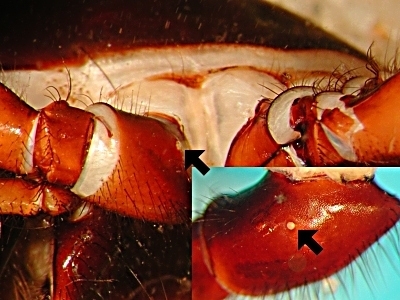
A pallid unsclerotised area, roughly circular in shape, on the posterior lower corner of the inner surface of the chelicerae of the males of some mygalomorph genera.
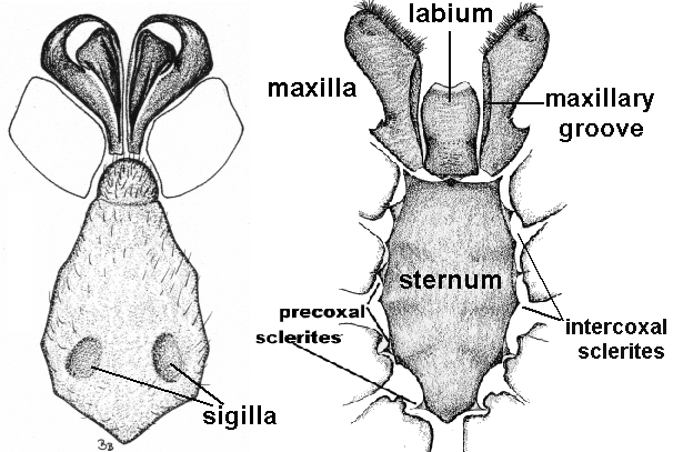
Sclerotised projections of the sternum that extend between the coxae. Sometimes the extensions join the sternum to the carapace; sometimes the sclerites are separate to the sternum.

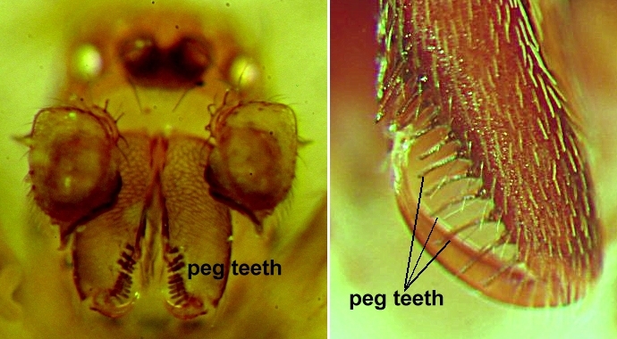
Short black studs or pegs on the ventral surface of the labium of mygalomorph spiders. In male mygalomorphs they may be tiny, pointed, incipient or absent. In female mygalomorphs they are rounded.

The unpaired structure behind the fangs, anterior to the sternum and in between the maxillae. The junction of the labium with the sternum is called the labiosternal junction, and is usually indicated by a groove. But in many araneomorph spiders, it is clearly joined by a flexible strip and is then termed 'free'. In cases where there is no flexible strip, the labium is termed 'fused'.

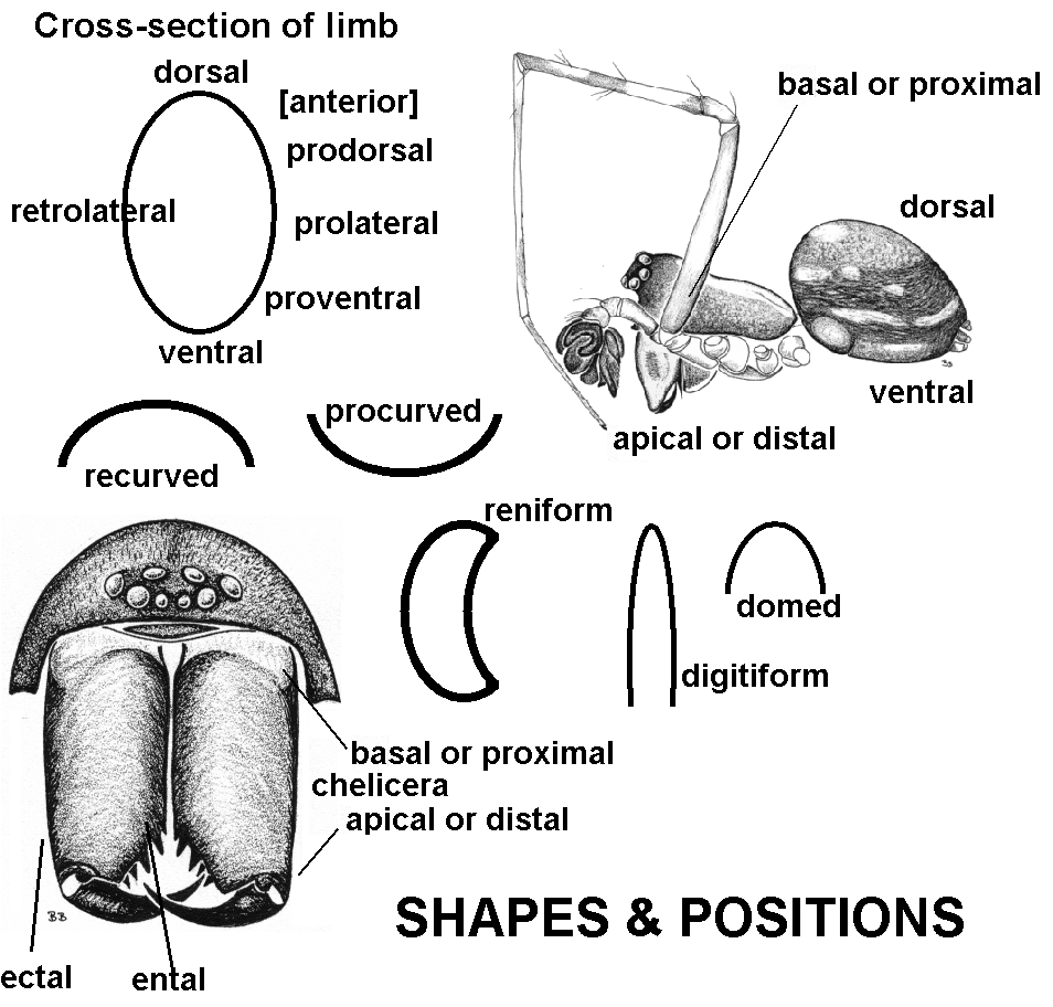
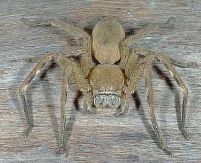
The legs are directed like those of a crab. The inner lateral surfaces of the femora (and other leg segments) face upwards, and femora often point laterally or posteriorly. In many groups, all four pairs of legs are 'turned' in this manner, such as family Sparassidae (huntsmen spiders). On the other hand, some Clubionidae have on the first two pairs of legs turned in this manner. Note that only araneomorph spiders have laterigrade legs.

See maxillary lyra.

The modified first segment (or coxa) of the palps (paired); and can be found just anterior to the sternum and posterior to the fanges. The maxillae are also called gnathocoxae or endites.

In mygalomorphs. A conical lobe on the maxillae that is partially defined by a groove posteriorly. The maxillae (palp coxae) are wider than long and almost transverse.
Short black studs or pegs on the ventral surface of the maxillae of mygalomorph spiders. In male mygalomorphs they may be tiny, pointed, incipient or absent. In female mygalomorphs they are rounded.
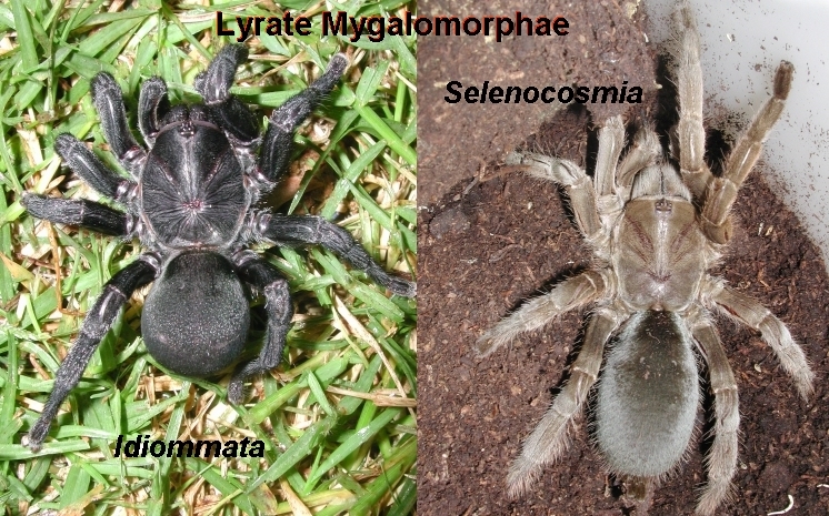
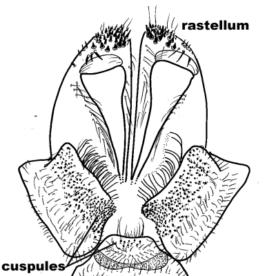
On closer examination (see inset in figure), the cuspules are socketed and have a characteristic surface pattern of finger-print like ridges. Cuspules are defined as 'densely packed' (left) when they are too numerous to count in 15 seconds. Densley packed cuspules only occur in Australian Tarantulas (Theraphosidae: Selenocosmiinae) and Funnelweb spiders (Hadronyche, Atrax).

Grooves occur in at least three locations on the maxillae: 1. Along the inner margin for the entire length of the maxilla, as in Lamponinae; 2. Across the maxillae giving the maxillae a saddle-like appearance, or a distinct groove around the middle of the maxillae; 3. a shallow diagonal groove beginning at the base or back of the maxilla and extending diagonally to the approximate centre of the maxilla, as in some Miturgidae.

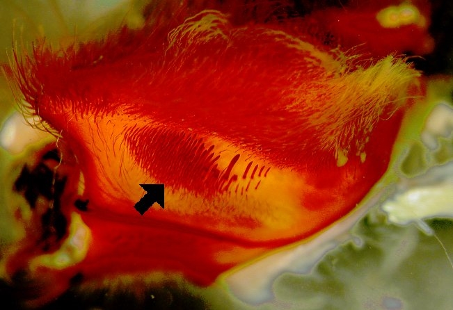
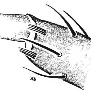
A transverse band of long thick spines on the anterior surface of the maxillae, that is directed down the face of the spider. This band of spines interacts with a line of fine 'pins' or 'strikers' located along the outer edge of the cheliceral furrow. The pins are believed to move across the spines on the lyra and create sound. In Selenocosmia, the spider makes an audible hiss when aggravated.
A line or band of short to minute spines or teeth on the anterior distal surface of the maxillae, i.e. the surface that lies in contact with the back of the chelicerae. It is best seen with the spider on its back and looking directly down on the maxillae - it will be visible as a black line or band. In araneomorph spiders, it is very rarely absent (e.g. Zodariidae), but on very small spiders it may be particularly difficult to see. The function of the serrula is unclear, but its position and structure indicate that it supports, or augments, the breaking up of prey during feeding.
The penultimate segment on the leg; the second most distal segment before the tarsus (and claws). The metatarsus is absent on the palps. See leg segments.

A notch in the margin of the trochanter segment (see leg segments), best visible when spider viewed from below and bending the femur back to vertical. The notches are either uniform narrow and deep (as found in many hunting spiders), or they are asymmetrical and triangular with deepest part of notch posteriorly. In the latter, the notches become shallower and more indistinct from coxa IV to coxa I. Their function is unclear, and notches are notably absent in the actively hunting Zodariidae and most Corinnidae.
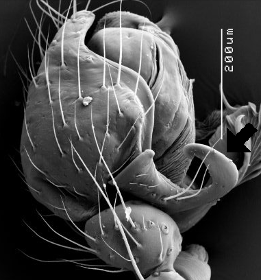
An extension of the unsclerotised portion of the tarsus between the claw base and the end of the tarsus.
The most anterior pair of leg-like appendages. The segments of the palps are the same as for the legs, except that the metatarsus is absent on the palps. The palp tarsus of females has only one claw, which is sometimes absent. The palp tarsus of adult males is modified into an intromittent sperm transfer organ. The claw has transformed into a complex structure usually with quite different shapes and processes between each species. See also cymbium, embolus, tegulum, and tibial apophysis.
A sclerotised process on the retrolateral edge of the male palp of entelegyne Araneomorphae. It is best developed in the Araneiodea, especially the Linyphiidae. The paracymbium is literally any process on the retrolateral side of the cymbium, and although the paracymbia may not have evolved from one kind, they are all scored here as one. Note that a spine (with a distinct socket, but not articulated) appears in this position in some Corinnidae: Castianierinae; and this spine may be difficult to distinguish from a paracymbium on smaller spiders.

When the action of the fang is down (or aligned with) the body (see fang action).
The fourth segment of the legs and palp (after coxa, trochanter and femur). It is a short segment between the long femur and long tibia. See leg segments.
The moult before becoming an adult. In male araneomorph spiders, the tarsus of the palp becomes swollen basally by the penultimate moult for the entire stage. In male mygalomorph spiders, the palp tibia becomes swollen only later in the penultimate stage. Soon after, that the cuticle appears to darken as the new cuticle and hairs begin to develop and become visible through the old cuticle.
Posterior lateral eyes.
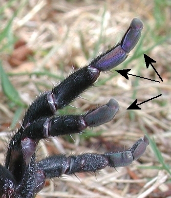
Posterior lateral spinnerets. PLS are always the biggest spinnerets in mygalomorph spiders, and are present on all. In araneomorph spiders, PLS are enlarged in the Hahniidae and Hersiliidae.

Posterior median eyes.
Posterior median spinnerets. Small in most mygalomorph spiders. Difficult to see in most araneomorph spiders because they are hidden by the ALS (anterior lateral spinnerets) and PLS (posterior lateral spinnerets). Most mygalomorphs retain PMS, but they are reduced or lost in some families, e.g. Barychelidae, Paratropididae, Idiopidae, Microstigmatidae). In araneomorphs, the PMS are considered to be used by females when producing the silk for the eggs sac, and hence PMS are sexually dimorphic.
Sclerotised plates or depressions located behind the entrance of the anterior book-lungs in some corinnids and lamponids. They may be separated from the scute that covers the book-lungs, or can be a lateral extension of it.
Triangular extensions of the sternum that insert into the base of the leg coxae; also called intracoxal processes.

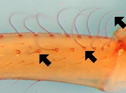
When present, it occurs on all leg tarsi and is sometimes more easily seen in the lateral view when using light microscopy as a pallid diagonal zone predistally on the tarsus. The crack is often not visible using SEM, presumably because the leg muscles harden and pull the fracture tight.
A group of similar closely-set setae, usually on a common base on the proventral and retroventral distal edges of the metatarsus. They are rare in spiders but, if present, will be present on metatarsi III and IV. They are present on all metatarsi in some Euagrinae mygalomorphs.
When viewed from dorsal angle, the eye row is bowed backwards so that the lateral edges are in front of the mid-point. Eye row curvature is measured by a line drawn through the centers of the eyes in that row. When viewd from front angle, this eye row curvature would be called downcurved.

The inner surface of the femora are almost vertical; and front legs are directed forwards. This is the most common and most widespread condition in spiders.
On the front side of the segment/structure. On the back legs, the prolateral side is the side facing away from the abdomen.

The prolateral, anterior or inner (mygalomorphs) margin on the cheliceral furrow.
On the lower half of the limb on the front side of the segment/structure. On the back legs, the proventral side is the side facing away from the abdomen.


Any modification of the distal edge of chelicerae above the fang that may facilitate digging and smoothing burrow walls. A rastellum may consist of a group of thick bristles or spines. They are set on a smoothly curving anterior surface of the chelicerae or on a raised mound. The rastellum is best seen when viewing the spider from below. If the setae on the rastellum are basally wide and have long slender stips, they are considered to be spine-like bristles. Whereas, spines are typically short and blunt, and often broken.
When viewed from dorsal angle, the eye row is bowed forwards so that the lateral edges are behind the midpoint. Eye row curvature is measured by a line drawn through the centers of the eyes. When viewed from front angle, this eye row curvature would be called upcurved.


This structure is readily visible with light microscopy on a medium-sized spider (e.g. a lycosid or a thomisid) as a pallid unsclerotised circle on the ental half of the retrolateral surface of coxa I. To observe this little window, the spider is best placed with the sternum facing upwards, then coxa I is held with one pair of forceps and rotated so that the retrolateral surface is clearly visible, and at the same time pull coxa II in a posterior direction to be clear of coxa I.
On the back side of the segment/structure. On the back legs, the retrolateral side is the side against the body.


The retrolateral, posterior or outer (mygalomorphs) margin of the cheliceral furrow.


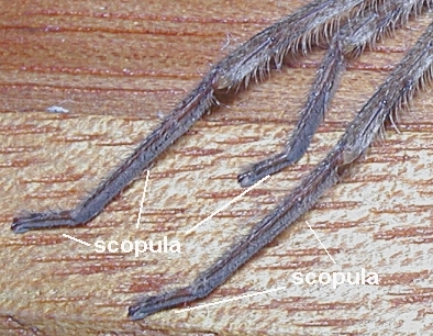
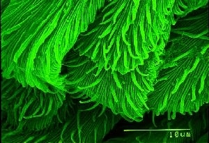
Dense hairs with enlarged tips (not always visible under stereo microscopy). The hairs arise at right angles to the cuticle surface. Scopula hairs are not under hydraulic control and do not extend if the leg is squeezed. The scopula along with the claw tufts allow the spider to climb smooth sloping and vertical surfaces. Using a scanning electron microscope, the scopula hairs are paddle-like and covered with finely divided filaments which act to increase the surface area and hence aid with adhesion. See also claw tufts.

A sclerotised, hard and often shiny portion of the cuticle of the abdomen; the hardness can be easily checked by applying gentle pressure to the suspect area with a pair of forceps.
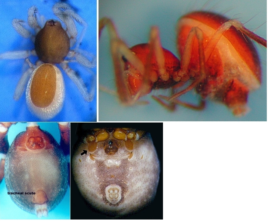
Images in figure: top row - dorsal scutes on the abdomen of Oonopidae (left) and Matilda (right); bottom row - tracheal scute on Corinnidae (left), and scutes on book-lung covers of Theridiidae: Hadrotarsinae.

With a saw-tooth edge. Thick spine-like setae on the ventral surface of tarsi IV of the Theridiidae have serrated edges.
See maxillary serrula.
See bristle(s), spine(s), hairs(s) or trichobothria.
Determination of the sex of most spiders is quite simple when the spiders are adult.
In adult males, the terminal segment of the palp (tarsus or cymbium) is transformed into a highly modified sclerotised structure (see image adut male) used for the transfer of sperm from the male genital pore via a tiny temporary sperm web.
In adult females, the situation depends of the group of spiders. In mygalomorphs and haplogyne araneomorph spiders, there is little or no special external indication of the reproductive structures. In contrast, in entelegyne araneomorph spiders, the external copulatory pore (gonopore) is marked by often highly sclerotised and complex ridges, plates and septa (epigyne) that guide the male palp embolus into the gonopore.
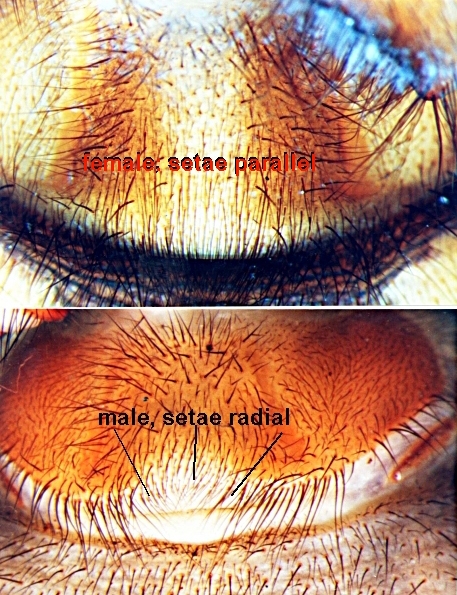
In all spiders, however, it is possible to establish the sex with high reliability simply by careful examination of the epigynal fold between the anterior book-lungs. In females, the setae around the midline of the genital area are parallel to the long axis of the spider and the fold is smoothly convex. In males (including subadult or juvenile males), the setae around the midline of the distal edge of the genital fold are radial; and the area is pallid and often medially indented thus interrupting the line of the smooth convex curve.
See also penultimate.

Oval or suborbicular (or rarely with irregular outline) depressions on the sternum of Mygalomorphae, and in the araneomorph family Filistatidae. Sigillae are paired (symmetrical down body midline) and represent the sites where large muscles attach to the endosternite.

Thick, strong, rigid, often ending abruptly, and usually straight rods of chitin, often with longitudinal ridges or fluting. They are socketed, and are raised and lowered under hydraulic pressure to the joint. Spines are usually few in number and considerabley thicker and sturdier than the surrounding bristles or setae. Spines may also be called macrosetae. Megaspines are immovable spines that are much thicker than common spines, and are used to lock open the female's fangs or to lock the base of the female's leg in order to attain a 'safe' mating position. Some megaspines occur on the palpal tibia of male mygalomorph spiders. See also bristle(s) and hairs(s).

A group of small, paired conical or finger-like structures at the posterior end or posterior ventral surface of the abdomen; represent the outlets for the silk glands.

The large ventral plate located in the middle of all the leg coxae to which muscles of the endosternite attach internally.

There are two different kinds. One is a regularly corrugated area against which another hard sclerotised structure is rubbed. This kind is diagnostic of Linyphiidae, in which it is situated on the outer edge of the chelicera.
The second is a group of special spines or hairs, against which another set of spines is strummed. See also maxillary lyra.
Shallow grooves under the eyes.
A low-set sensory structure positioned dorsally on the leg tarsi; usually distal of the most distal trichobothria. The tarsal organ is a chemosensory structure. The tarsal rod is a homologue of the tarsal organ.
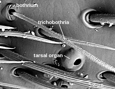

A long tarsal rod, set at approximately 40-50 degrees to the cuticle surface, is present at approximately the mid-point (or just basal to it) on the leg tarsi in some araneomorph and mygalomorph spiders. The rod is not present on the palp tarsi of either sex. The tarsal rod is a homologue of the tarsal organ.


The large basal portion of the adult male palp on which the other palp sclerites are located (embolus, conductor, median apophysis). The tegulum is attached to the bulb through the subtegulum, and these are joined by flexible unsclerotised tissue.


Thick paddle-shaped hairs found below the claws and often mistaken for claw tufts. Unlike tufts, they are not on separate pads and are not moveable with hydraulic pressure. See also claw tufts.


The fifth segment of the legs and palp (counting from the body out along leg). The segment after the short patella. See legs segments.
A sclerotised process on the retrolateral surface of the adult male palp tibia. The sclerotised process is an extension of the cuticle and not a separate or articulated spine. Also called RTA or retrolateral tibial apophysis.

A fine discontinuity (resembling a crack or thinning) that occurs on the leg tibiae of male Lycosidae. It is very close the base of the tibia segment, and there is often a ventral or lateral spine on its ditsal side. The suture is visible with a dissecting microscope as a discolouration or a shallow groove. It is often easily seen on the retrolateral side of the tibia.
The tracheal spiracle is considered to be the external indication of the vestige(s) of the posterior book-lungs; and is often visible as a subtle transverse slit ventrally on the abdomen. In most araneomorphs, it is usually narrow and closer to the spinnerets. In some spiders it is wide (e.g. Cyatholopidae), situated as far from the spinnerets as the epigastric furrow (e.g. Hahniidae, Anyphaenidae), and in some haplogyne spiders the tracheal spiracle is paired and situated just posterior to the book-lung aperatures. Mygalomorph spiders lack tracheal spiracles.

Fine, often very long, hairs on the upper surface of the tibia, metatarsus and tarsus (and rarely on the retrolateral surface of the dorsal femora, e.g. Tetragnathidae and Uloboridae), where they usually form lines. In some groups, the trichobothria may be reduced to just one present on the metatarsi, e.g. the Linyphiidae; and rarely absent entirely on the tarsi. Trichobothria are easily moved when the legs are moved in alcohol or in the wind if specimen is dry.
Trichobothria can be clavate on mygalomorphs.


The second segment of the legs and palp (counting from the body out along the leg). The trochanters are mostly only visible when spider is viewed from below, and usually a short collar-like ring segement between the coxa and the femur. Many hunting spiders have a distal notch in the margin of the trochanter only visible when viewed from below.

See claw tufts.
When viewed from the front angle, the lateral edges of the eye row are curved upwards. Eye row curvature is measured by a line drawn through the centers of the eyes in that row.